An exact 3D replica of a person's heart, printed thanks to a soft and flexible robotic model capable of contracting like the original. A successful experiment whose result is published in the journal Science Robotics by a group of engineers from MIT (Mit) coordinated by Ellen T. Roche. The 3D heart will help doctors study the anatomy and organ function of individual patients, so they can tailor specific treatments.
The heart in 3D
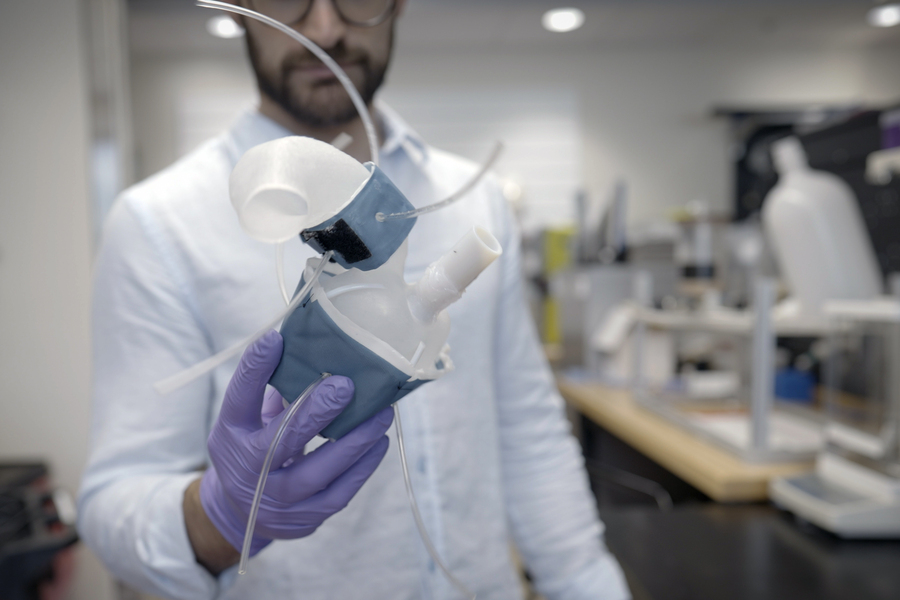
The first author of the study is Luca Rosalia, a young researcher from Catania who trained between Great Britain, Singapore and the United States and who developed the robotic heart project while he was locked in his room on the university campus during the lockdown in March 2020. The robotic model is created starting from the images of the organ collected for diagnostic purposes: these are converted into a digital model on the computer which is then 3D printed using a special polymer-based ink.
In this way a soft and flexible shell is obtained which has the same shape as the patient's heart. The same can also be done for the aorta, the main artery that carries blood from the heart to the rest of the body. To simulate contraction, the 3D printed heart and aorta are lined with sheaths, similar to the cuffs on blood pressure monitors. They are connected to a pneumatic system with which air is rhythmically introduced to induce contraction. The constriction can be adjusted to simulate aortic stenosis as well. According to MIT researchers, 3D heart replication will help doctors choose the best model of artificial valve to implant in the future. It could also be used in research laboratories and by the biomedical industry as a platform for testing new therapies.
(Source: Melanie Gonick, MIT)